Role of Recombinant Proteins for Treating Rheumatoid Arthritis
-
 Soleimani Sasani, Mahboubeh
1-Shahid Beheshti University, 2-Department of Biotechnology Engineering, Faculty of Medical Engineering, Shahab Danesh University, Qom, Iran, Tel: +98 9120319531 Fax: +98 25 32317448; E-mail: mah.slmn@yahoo.com
Soleimani Sasani, Mahboubeh
1-Shahid Beheshti University, 2-Department of Biotechnology Engineering, Faculty of Medical Engineering, Shahab Danesh University, Qom, Iran, Tel: +98 9120319531 Fax: +98 25 32317448; E-mail: mah.slmn@yahoo.com
-
Moradi , Yeganeh
-
Department of Biotechnology Engineering, Faculty of Medical Engineering, Shahab Danesh University, Qom, Iran
Abstract: Rheumatoid Arthritis (RA) is an autoimmune disease and chronic inflammatory disorder that affects joints and causes inflammation, pain, stiffness, and eventually progressive joint destruction. Approximately 1% of the world's population is estimated to suffer from RA, and if this disease is left untreated, it can lead to severe disability. Despite all the efforts and advances made by professionals in the field, there is currently no definitive treatment for RA, and most treatment strategies are aimed at relieving symptoms and improving patients' quality of life. One of the most promising current approaches is the use of recombinant proteins that target specific signaling pathways involved in the development of RA to alleviate symptoms and slow the progression of the disease. This article discusses the genetic and immunological factors that influence the development of RA, recombinant proteins, methods of using these proteins, approved drugs, and side effects associated with treating RA.
Introduction :
Introduction and description of the problem: Rheumatoid Arthritis (RA) is a chronic inflammatory disease that affects synovial joints and leads to pain, stiffness, and reduced mobility. Although the exact cause of this disease is not fully known, it appears to be an autoimmune disease in which the immune system mistakenly attacks the body's tissues. This problem is the result of a combination of genetic factors, the environment, and the immune system. RA is a complex disease involving both resident cells and infiltrating cells in membrane tissue 1. In this disease, significant amounts of new vessels are formed in the membrane tissue, facilitating invasion by lymphocytes and monocytes and transforming a cell-free, loose cavity membrane into an abnormal, tumor-like invasive tissue. Micro vessels proliferate, forming straight vessels and branching regularly 1. Studies have shown that the synovial fluid in patients with RA is highly inflamed in all joints and expresses much inflammatory genes 2. The pathophysiology of RA is heterogeneous and includes defects in the innate and adaptive immune systems, genetic and environmental factors, autoantibodies, cellular changes, signaling pathways, and metabolism 1. Understanding the role of individual variations in the cellular and molecular mechanisms associated with RA will significantly improve clinical care and patient out-comes. Individualized responses to standard therapy are observed in RA because of pathophysiological heterogeneity, ultimately leading to poor overall prognosis 2. Key cellular and molecular findings in RA include angiogenesis, B cells, fibroblasts, reduced oxygen levels, synovial tissue, and T cells. Researchers are investigating altered metabolic pathways underlying synovial inflammation in RA 1. The molecular and cellular heterogeneity of RA is a hot topic, and understanding the underlying mechanisms may lead to therapeutic intervention 3. RA is currently treated worldwide to reduce inflammation, relieve pain, and slow the progression of joint damage. Conventional treatments include nonsteroidal anti-inflammatory drugs, disease-modifying antirheumatic drugs, and biological agents, and in recent years, recombinant proteins have emerged as promising biological agents that can target specific proteins or cells involved in the inflammatory response 4. Recombinant proteins are synthetic proteins that are produced in the laboratory using genetic engineering techniques. These proteins can mimic the functions of natural endogenous proteins, such as cytokines, growth factors, and antibodies. In the case of RA, recombinant proteins can be used to target specific proteins or cells involved in the inflammatory response 5. Abatacept, for example, is a recombinant protein that binds to T cells and plays a key role in the immune response and prevents their activation. By inhibiting the activation of T cells, this protein can reduce inflammation, slow the progression of joint damage, and be effective for treating RA 6.
Factors Influencing the Development of RA: In addition to environmental factors such as smoking, infections, and exposure to certain occupational hazards 7, several genetic factors, such as PTPN22, STAT4, TRAF1, and TNFAIP3, have been identified as being involved in the pathogenesis of rheumatoid arthritis 8. However, the best-known genetic risk factor is the HLA-DRB1 gene, which codes for a protein involved in antigen presentation on T cells. Certain variants of the HLA-DRB1 gene, known as shared epitope alleles, are also strongly associated with an increased risk of developing this disease 9. Because RA is classified as an autoimmune disease, immunological factors are among the most potent factors for the onset and exacerbation of this disease. In autoimmune diseases, the immune system mistakenly attacks the body’s tissues. In this disease, the immune system targets synovial fluid tissue, leading to chronic inflammation and joint damage. The exact mechanisms of immune system activation in RA are not yet fully understood, but several immunological factors are involved 10.
T cells: T cells play an important role in the pathogenesis of RA. In particular, CD4+T cells, also known as helper T cells, are involved in the activation of other immune cells, such as B cells and macrophages, and the production of proinflammatory cytokines, such as TNF-α and IL-17. In addition, the number and function of regulatory T cells, which contribute to the suppression of the immune response, are reduced in patients with RA 11.
In RA, the infiltration of T cells into the synovial membrane triggers the activation of macrophages and synovial fibroblasts, transforming them into tissue-destructive effector cells. In this disease, multiple T cell activation pathways are involved, which are combinations of HLA class II molecules. Aberrant pathways of T-cell activation, differentiation, and persistence play a key role in RA, along with disease-associated variants such as HLA-DRB1, PTPN22, CTLA4, IL2RA, IL2RB, and STAT4 12. In addition, in patients with RA, it has been observed that clonally expanded CD4+ T cells lose expression of the CD28 molecule and gain expression of perforin and granzyme. Consequently, the functional profile of expanded CD4(+)CD28 null T cells is fundamentally skewed towards causing tissue damage and vascular injury 13.
B cells: As RA progresses, the chronic inflammatory reaction in the synovial membrane causes the migration of monocytes, T, and B cells into the stroma under the synovial layer. The B cells in the synovial tissue are activated by autoantigens. This leads to the formation of aberrant germinal centers, the formation of plasma cells, and subsequently the activation and differentiation of B cells. With the formation of plasma cells, autoantibodies with high affinity are secreted into the synovial tissue. The formation of the antigen-antibody complex, the activation of the complement cascade, and the stimulation of macrophages lead to further destruction of the joints. B cells play an essential role in the activation of synovial T cells and the triggering of cytokine secretion. Cytokines of the TNF family, such as TNF-α or lymphotoxin as well as IL-6, can play an important role 14. These cytokines not only intensify the inflammatory process but are also very important for the ontogenesis of lymphoid organs.
B cells have several functions in the immune response. They are important as precursor cells for plasma cells and thus for antibody production 15. In addition, B cells are also regulatory cells that can influence the disease process through the production of cytokines. Another function of B cells is the presentation of antigens and thus the activation of T cells. In around 10% of patients, large cell clusters can be seen in a follicle-like structure containing CD20+B lymphocytes. At the center of these large cell clusters is a network-like structure formed by Follicular Dendritic Cells (FDC). The task of these dendritic cells is to present B-cell antigens. The FDC carries the antigen on its surface in the form of antigen-antibody complexes. The antigen-antibody complex binds via the complement receptor and the Fc receptor and is thus presented to the B cells. This FDC-like network structure can only form in the presence of lymphotoxin-positive B cells. B cells receive important survival signals from FDCs. FDCs also play an important role in the T cell-dependent immune response. In the germinal center reaction, FDCs support the proliferation of B cells. They are responsible for ensuring that only B cells with a high-affinity antigen receptor differentiate into plasma or memory cells. It has also been discussed that fibroblasts, which are activated by chronic inflammatory processes in the synovial tissue, differentiate into FDC. Interestingly, FDC networks and the resulting formation of aberrant germinal centers have also been described in other autoimmune diseases with local chronic inflammation 16,17.
Cytokines: Cytokines are small proteins that play a role in regulating the immune response. In RA, several cytokines are involved in the pathogenesis of the disease, including TNF-α, IL-1, IL-6, IL-17, and IL-23. These cytokines activate immune cells and stimulate vascular function. The generation and production of matrix metalloproteinases increase inflammation and joint damage 18.
Biomarkers of RA: Biomarkers are measurable indicators that can provide information about the presence, severity, and progression of disease. In recent years, significant progress has been made in the identification and validation of biomarkers for RA 19. These biomarkers are classified to following items:
Diagnostic biomarkers: Several biomarkers have been identified that can assist in the diagnosis of RA. These include autoantibodies such as rheumatoid factor and antibodies against cyclic citrullinated peptides, which are present in approximately 70-80% of patients with RA 20. Other biomarkers that have been proposed for the diagnosis of RA include cytokines, such as IL-6 and IL-33, and chemokines, such as CXCL13 21,22.
Prognostic biomarkers: Prognostic biomarkers are indicators that can predict the severity and progression of RA. Several markers, including increased levels of acute-phase reactants, such as C-Reactive Protein (CRP), the Erythrocyte Sedimentation Rate (ESR); and autoantibodies, such as anti-CCP antibodies, are associated with a worse prognosis in patients with RA 23,24. Other biomarkers that have been proposed as prognostic indicators in RA include cytokines such as IL-6 and TNF-α 25 and chemokines such as CXCL10 25.
Predictive biomarkers: Prognostic markers can predict the response to a specific treatment in patients with RA especially in the period of biological medicines. Several markers, such as TNF-α inhibitors, indicate the response to biological agents. These autoantibodies include autoantibodies, such as rheumatoid factor and anti-CCP antibodies, and cytokines, such as IL-6 and IL-10 26,27. Other biomarkers that have been proposed to predict the response to biological agents in patients with RA include MRI with contrast agents and ultrasound 28,29.
Currently, predictive biomarkers are being investigated for each biological drug .The biomarkers that predict a better response to rituximab treatment include lower levels of type I interferon (IFN-γ), reduced serum levels of B-cell activating factor (BAFF) or B lymphocyte stimulator (BLyS), and a favorable Fcγ receptor III (FcγRIII) genotype 30. Predictors in patients treated with tocilizumab are increase the baseline ESR of >30 mm/hr and baseline CRP levels of >10 mg/L, and the presence of extra-articular disease manifestations 31.
Monitoring of biomarkers: Monitoring biomarkers are indicators that can be used to assess the response to treatment and disease activity in patients with RA. Several markers, including acute-phase reactants such as CRP and ESR, as well as cytokines such as IL-6 and TNF-α, are useful for monitoring disease activity in patients with RA. Other biomarkers that have been proposed for monitoring disease activity in RA include imaging techniques such as ultrasound and MRI, as well as evaluating indices such as the disease activity score, the simplified disease activity index, and the clinical disease activity index 32-34.
Recombinant proteins used to treat RA: The use of recombinant proteins has emerged as a promising treatment for RA because they can specifically target the inflammatory pathways involved in the disease. However, it is always important to note that the dose and frequency of these treatments may be adjusted on the basis of the patient's response and disease activity, as well as other factors such as side effects and drug interactions. Therefore, patients who receive these treatments should be carefully monitored by a healthcare provider to ensure optimal dosage and frequency 4.
Tumor necrosis factor inhibitors: TNF inhibitors are a class of recombinant proteins that bind to and neutralize TNF, a proinflammatory cytokine that plays a crucial role in the development of RA. Several TNF inhibitors 18, including infliximab, adalimumab, etanercept, certolizumab pegol, and golimumab, are currently available. Among these drugs, infliximab, adalimumab, and etanercept are the three TNF inhibitors currently approved for the treatment of this disease 4,35,36. These agents are effective at reducing disease activity, improving joint function, slowing radiographic progression, alleviating joint symptoms, and reducing inflammation and disease progression in patients with RA. Unfortunately, these treatments are also associated with an increased risk of infection and other side effects, such as injection site reactions 4.
Infliximab is a chimeric murine variable region/ human IgG1. It is a monoclonal antibody (mAb) biologically classified as human Tumor Necrosis Factor-α (TNFα). the protein chemical formula is C6428H9912 N1694O1987S46 with a weight near 145 kDa 37. After infliximab injection, this drug shows a good relationship between the administered dose and the maximum amount in blood. In patients with RA, the maximum infliximab concentration after a single dose injection of 5 mg/kg, 10 mg/kg, and 20 mg/kg was 192±51 µg/ml, 427±106 µg/ml, and 907±183 µg/ml, respectively 38.
Adalimumab is a mAb composed of two kappa light chains each having a molecular weight of approximately 24 kDa and two IgG1z heavy chain, each with a molecular weight of approximately 49 kDa with a protein formula of C6428H9912N1694O1987S46 and its molecular weight is 145 kDa 39,40. After a single subcutaneous injection of 40 mg adalimumab into healthy adults, peak plasma concentration and time to peak were 4.7±1.6 μg/ml and 131±56 hr, respectively. The mean bioavailability of adalimumab after a single 40 mg subcutaneous dose was estimated to be 64% from three clinical studies. After a single injection, the pharmacokinetics of adalimumab exhibited a more linear pattern between 0.5 and 10.0 mg/kg after a single intravenous dose 41.
Full-length IgG1 antibodies, such as infliximab and adalimumab, can inhibit TNFα-producing cells by forming a 1:2 complex with the transmembrane TNFα trimer, causing apoptosis and cell cycle G0/G1 arrest 39. Etanercept is a dimeric fusion protein produced by recombinant DNA technology, that combines two naturally occurring soluble human 75 kDa TNF receptors linked to an Fc portion of IgG1. Etanercept is a complex molecule that contains 6 N-glycans, up to 14 O-glycans, and 29 disulfide bridge structures 42,43. This is a 51234.9 Da molecule protein with chemical formula C2224H3475N621O698S36 44. Pharmacokinetic modeling in adults with RA, Ankylosing Spondylitis (AS) and healthy individuals showed a subcutaneous absorption of 56.9% and a Ka of 0.0223/hr. Another sample of pediatric patients with Juvenile Idiopathic Arthritis (JIA) showed an increase in Ka of 0.05/hr with a high mean interindividual variability of up to 215%. After a single 25 mg subcutaneous injection of Enbrel, maximum concentration (Cmax) is 1.1 µg/L and time to maximum (Tmax) of 69 hours. According to reports, when using a dose of 25 mg twice a week in adult RA, Cmax after repeated administration is 2.4 µg/L; Cmax in JIA pediatric patients when the dose is 0.4 mg/kg twice weekly is 2.1 µg/L 45,46.
Interleukin-6 (IL-6) inhibitors: IL-6 inhibitors are a class of recombinant proteins approved for the treatment of RA. IL-6 is a cytokine that increases RA and contributes to inflammation and joint destruction 47. Tocilizumab and sarilumab are two IL-6 inhibitors currently approved for the treatment of RA 4. These drugs, which are recombinant human monoclonal antibodies, bind to the IL-6 receptor and block its signaling pathway 47, ultimately reducing its activity. Disease, improving joint function, and slowing radiographic progression in patients with this condition are effective in improving joint symptoms by reducing inflammation. However, these drugs are still associated with an increased risk of infections and other side effects, such as elevated liver enzymes and gastrointestinal perforation 4,47.
Tocilizumab is a humanized recombinant mAb of the IgG1 kappa subclass produced by recombinant DNA technology. The antibody is composed of two heavy chains and two light chains with 12 intrachain and four interchain disulfide bonds, and with a total mass of 149 kDa (Sheppard et al, 2017). The chemical formula of TCZ is C6428H9976N1720O2018S42 48. Tocilizumab inhibits IL-6 classical and trans signaling pathways by binding to membrane-bound IL-6R (mIL-6R) and soluble IL-6R (sIL-6R) 47. Tocilizumab binds to the receptor, preventing it from binding to IL-6. The resulting tocilizumab–receptor complex is biologically inactive because it cannot affect the dimerization of the gp130 molecules. In the absence of dimerization, the IL-6 signaling is completely blocked 47. At a subcutaneous dose of 162 mg every week Cmax is 51.3±23.2 µg/ml and Area under the ROC Curve (AUC) is 8254±3833 µg*hr/ml. Administration of the same dose every 2 weeks resulted in a Cmax of 13±8.3 µg/ml and an AUC of 3460±2530 µg*hr/ml. At a dose of 162 mg administered subcutaneously every 4 weeks, Cmax was 154±42 µg/ml and AUC was of 39216±14304 µg*hr/ ml 49.
Sarilumab is a mAb with protein structure C6388 H9918N1718O1998S44 and a molecular weight of approximately 150 kDa. Sarilumab is well absorbed after subcutaneous injection in patients with RA and reaches maximum levels in the blood after 2-4 days. For the 150 mg dose every 2 weeks, the AUC, Cmin, and Cmax values of sarilumab were 202±120 mg·day/L, 6.35±7.54 mg/L, and 20.0±9.20 mg/L, respectively. the AUC, Cmin, and Cmax values of sarilumab were 395±207 mg·day/L, 16.5±14.1 mg/L, and 35.6±15.2 mg/L, respectively. For the 200 mg every two weeks dosage regimen 50,51. Sarilumab is a covalent heterotetrametric consisting of two disulfide-linked heavy chains covalently linked to a kappa light chain. The heavy chain consists of a continuous IgG1 constant region with a single an N-linked glycosylation site in the Fc portion of the molecule. Complementation Determining Regions (CDRs) in the variable domains of both light and heavy chains combine to form the binding site for IL-6R. As an IgG1 molecule, sarilumab mediates Fc effector activity by binding to IL-6Ra, and it rapidly binding to FcγRI, FcγRIIa, FCγRIIb, FcγRIIIa, and FcγRIIIB 52.
Interleukin-1 inhibitor: IL-1 is another pro-inflammatory cytokine involved in the pathogenesis of RA. Anakinra is a recombinant human IL-1 receptor antagonist that blocks the activity. It is effective in reducing disease activity and improving joint mobility in RA patients 53. Its use is limited due to need for daily subcutaneous injection and the potential of injection sites 36.
Anakinra is an interleukin product with the chemical formula C759H1186N208O232S10 and a molecular weight of 173 kDa. Anakinra is a recombinant human interleukin-1 (IL-1) receptor antagonist (IL-1Ra) containing 153 amino acid residues. Unlike native human IL-1Ra, anakinra has an additional methionine residue at its amino terminus. It also differs from the human protein in that it is produced in Escherichia coli, therefore is not glycosylated. By binding to the IL-1 receptor, the drug competes fir IL-1α and β and inhibits its activity 54,55.
Targeted B-cell therapies: B cells play a crucial role in the development of RA by producing auto antibodies and cytokines that lead to joint inflammation and damage. B-cell depletion therapy is a therapeutic strategy that targets B cells of the immune system that produce autoantibodies in patients with RA. Rituximab is a recombinant chimeric mAb that targets CD20, a B-cell surface antigen, and depletes B cells from the circulation. Rituximab is currently the only approved B-cell depletion treatment for RA 36. It effectively reduces disease activity, improves joint function, decreases inflammation, and slows radiographic disease progression. Improvement of physical performance in patients with RA is effective 56. However, it is associated with an increased risk of infections and other side effects, such as infusion reactions and hepatitis B virus reactivation 56.
Rituximab is a chimeric mouse/human mAb with the chemical formula C6416H9874N1688O1987S44 and a molecular weight of 144 kDa. There are 451 amino acids residues in the heavy chain of Rituximab and 214 amino-acid residues in the light chain. The heavy and light chains are connected by a disulfide (S-S) bond. The heavy chains are connected by two S-S linkages and an additional 12 cysteine bridges in the molecule. Both the LC and HC of rituximab contain N-terminal glutamine (Q1) and form N-terminal pyroglutame. An important change that occurs in recombinant mAbs is the cleavage of the C-terminal lysine 57. Rituximab binds to the CD20 antigen.
T cells: Blockers of T-cell stimulation are another class of recombinant proteins approved for the treatment of RA. T cells are immune cells that play a crucial role in the development of RA by promoting inflammation and joint damage. Abatacept is currently the only approved T-cell costimulatory blocker for treating RA 6. It improves joint symptoms, slows disease progression, and eliminates the need for other medications. However, it is still associated with an increased risk of infection and other side effects 36.
Abatacept is a recombinant fusion protein that combines the cell surface marker CTLA-4 with a fragment of immunoglobulin G. It links the extracellular domain of human cytotoxic T-lymphocyte-associated antigen 4 (CTLA-4) to the modified Fc portion (hinge, CH2, and CH3 domains) of human immunoglobulin G1 (IgG1). Abatacept is a glycosylated fusion protein with a molecular weight of 92 kDa. It consists of two homologous polypeptide chains, each possessing 357 amino acids, and forms a homodimer structurally. It is constructed using recombinant DNA technology in mammalian CHO cells.
Administration and side effects of approved recombinant proteins for rheumatoid arthritis: The revolution created by recombinant proteins in providing targeted and effective therapies to reduce inflammation and treat RA cannot be ignored. TNF inhibitors, IL-6 inhibitors, IL-1 inhibitors, and B- and T-cell-targeted therapies have been proven to be effective in reducing disease activity. Improving joint function and slowing radiographic progression are effective in patients with RA. However, these factors are associated with significant costs and potential side effects. Therefore, it is important to consider the method of consumption and develop strategies to control the side effects of these drugs. In addition to cost, careful monitoring and management of side effects are essential to ensure the safety and effectiveness of these treatments. The typical administration routes and side effects of these drugs are presented in table 1 5.
Conclusion :
Rheumatoid Arthritis (RA) is a complex autoimmune disease involving the interplay of genetic, environmental, and immune factors. Significant progress in understanding the cellular and molecular basis of RA has led to the development of more targeted and effective treatments. Current treatments for rheumatoid arthritis include Disease-Modifying Antirheumatic Drugs (DMARDs), biological agents, and small molecule inhibitors that target specific cells or cytokines involved in disease pathogenesis 2. However, future directions for research on rheumatoid arthritis include identifying new therapeutic targets and developing personalized therapies based on an individual’s genetic and immune profile. Therefore, several recombinant proteins, such as TNF-α inhibitors, IL-1Ra, abatacept, and rituximab, are available for the treatment of RA. These proteins have been proven to be effective in reducing the signs and symptoms of this disease. These drugs have revolutionized the treatment of rheumatoid arthritis by offering targeted therapies that can decrease inflammation and slow the progression of joint damage. However, as with any other treatment, careful monitoring and management of side effects are necessary to ensure the safety and effectiveness of these treatments 58. It is hoped that through further effective research conducted by experts in this field, the adverse effects of these drugs will be minimized, leading to self-sufficiency and independence in their production.
Acknowledgement :
This review article is written based on the opinion of the corresponding author and coworker of the second author. We are responsible for the content and any remaining errors, omissions, and inaccuracies.
Conflict of Interest :
The authors declare no conflicts of interest associated with this manuscript.
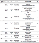
Table 1. Recombinant agents, methods of action, prescriptions, and side effects of recombinant proteins for the treatment of rheumatoid arthritis
|
|