The Effect of Biopsy During Precompacted Morula Stage on Post Vitrification Development of Blastocyst Derived Bovine Embryos
-
 Shirazi, Abolfazl
D.V.M., Ph.D., Research Institute of Animal Embryo Technology, Shahrekord University, Shahrekord, Iran, P. O. Box: 115, Tel: +98 381 4421626 Fax: +98 381 4424412 E-mail: shiraziabbas@yahoo.com, a.shirazi@avicenna.ac.ir
Shirazi, Abolfazl
D.V.M., Ph.D., Research Institute of Animal Embryo Technology, Shahrekord University, Shahrekord, Iran, P. O. Box: 115, Tel: +98 381 4421626 Fax: +98 381 4424412 E-mail: shiraziabbas@yahoo.com, a.shirazi@avicenna.ac.ir
-
Research Institute of Animal Embryo Technology, Shahrekord University, Shahrekord, Iran
-
Reproductive Biotechnology Research Center, Avicenna Research Institute, ACECR, Tehran, Iran
-
Borjian, Sara
-
Reproductive Biotechnology Research Center, Avicenna Research Institute, ACECR, Tehran, Iran
-
Ahmadi, Ebrahim
-
Research Institute of Animal Embryo Technology, Shahrekord University, Shahrekord, Iran
-
Nazari, Hassan
-
Research Institute of Animal Embryo Technology, Shahrekord University, Shahrekord, Iran
-
Heidari, Banafsheh
-
Reproductive Biotechnology Research Center, Avicenna Research Institute, ACECR, Tehran, Iran
Abstract: Improvements on embryo micromanipulation techniques led to the use of embryo biopsy in commercial embryo transfer programs for genetic analysis of preimplantation bovine embryos. The aim of this study was to evaluate the quality of bovine blastocyst derived from embryos biopsied at different precompacted morulae stages by assessment of cryosurvivability of the resulting blastocysts. The in vitro produced bovine embryos were subjected to biopsy at days 2, 3, and 4 post-insemination with different cell numbers (4 to 16-cells). Embryo cell biopsy was carried out in a 100 ?l drop of H-SOF following pronase drilling by aspiration of one blastomere. The biopsied embryos were then cultured in SOFaaBSA co-cultured with oviduct cells-monolayer until blastocyst formation. The blastocysts were cryopreserved at room tempera-ture after exposure of equilibration (glycerol 1.4 M for 5 min and then glycerol 1.4 M and ethylene glycol 3.6 M for 5 min) and vitrification solutions (3.4 M glycerol and 4.6 M ethylene glycol). The blastocysts were loaded into the center of 0.25 ml straws separated by air bubbles from 2 columns of sucrose 0.5 M and plunged immediately into liquid nitrogen. There was no significant difference in cryosurvivability of vitrified-warmed blastocysts derived form biopsied embryos at different pre-compacted morula stages. The quality of biopsy derived blastocysts was identical to that of non-biopsy derived ones in terms of post vitrifcation survival and hatching rates. In conclusion there was no preference between different times of embryo biopsy at precompacted morula stages in term of cryosurvivability of biopsy derived bovine blastocysts.
Introduction :
In transgenic technology, in order to pre select offspring harboring exogenous DNA and in farm animal industry, preselection of offspring sex screened by PCR (1, 2), the microsurgical sampling, so-called biopsy of blastomeres from the embryos is required.
Storage of biopsied embryos until the results of genotyping determination and the proper recipients are available is crucially needed (3 - 5).
These circumstances have challenged researchers to establish and evaluate the long term storage methods that render biopsied embryos viable and competent after transfer to recipients. While it is claimed that the biopsy procedure has no harmful influences on the viability of fresh embryos, the developmental capacity of biopsied embryos following transfer is considerably reduced when they are cryopreserved (6, 7).
It has been shown that among the preimplantation mouse embryos biopsied at the 4-cell, 8-cell and morula stages, the biopsy has the least impact on developmental potential in vitro and in vivo when performed at the 8-cell stage (8).
The in vitro viability of manipulated or vitrified sheep embryos was significantly lower at precompacted morula and compacted morula stages than intact embryos at the same stages. No differences, however, were found at the blastocyst stage. Moreover, the in vitro survival rate of precompacted morula which were manipulated and immediately vitrified was lower than in those manipulated and, after a temporary period of culture, vitrified at blastocyst stage (9). It has also been reported that short-term in vitro culturing after microsurgical biopsy prior to cryopreservation has improved the post –warming survival rate of bovine embryos (10).
Considering the beneficial effect of temporary period of culture between embryo biopsy and cryopreservation on post warming survival rate of embryos and considering the cryotolerance of In Vitro Produced (IVP) embryos as a criteria to evaluate the quality of IVP embryos, this study was conducted to evaluate the effect of age and cell number of embryos at the time of biopsy on the quality of biopsy derived blastocysts.
Materials and Methods :
Except where otherwise indicated, all chemicals were obtained from the Sigma (St. Louis, MO, USA).
In vitro embryo production
The production of bovine embryo was as previously described (11). Briefly, all visible ovarian follicles with a diameter of 2 to 8 mm were aspirated using gentle vacuum (30 mm Hg) and released into the preincubated hepes-TCM, supplemented with penicillin and streptomycin and 50 IU/ml heparin.
The Cumulus–Oocyte Complexes (COCs) with at least 3 layers of cumulus cells and oocytes with a uniform granulated cytoplasm,
were selected for the experiments. The selected COCs were in vitro matured in TCM199 supplemented with 10% FBS (Fetal Bovine Serum, Gibco 10270), 0.02 mg/ml cysteamine and 0.1 IU/ml FSH for 24 hr in 5% CO2 in air at 39 °C.
The motile spermatozoa were obtained by centrifugation of frozen–thawed semen on a discontinuous Percoll density gradient (1 ml 40% Percoll over 1 ml 90% Percoll) at 700×g for 20 min. In vitro-matured cumulus-oocyte complexes were co-cultured with motile spermatozoa at 1 × 10⁶ spermatozoa/ ml in TALP medium supplemented with 6 mg/ml BSA, 10 µg/ml heparin, and 0.3 mM sodium pyruvate for 22-24 hr at 39 ºC in 5% CO2 in air.
After fertilization, presumptive zygotes were mechanically denuded of their cumulus cells and cultured in SOFaaBSA co-cultured with oviduct cells-monolayer (SOF-OCM) under mineral oil in maximum humidified atmosphere with 5% CO2. Cell sampling was performed at 2, 3, and 4 day post insemination of cleaved embryos.
Embryo biopsy
The embryos at days 2, 3, and 4 post-insemination with different cell numbers (4 to 16-cells) irrespective of grade, were transferred into the manipulation drop (HEPES-SOF) and subjected to biopsy using Narishige micromanipulators (Japan) in conjunction with an inverted microscope with Nomarsky optics (IX71 Olympus, Tokyo, Japan). While the embryo was immobilized by suction with a holding pipette, a drilling pipette (internal diameter 22 µm) was placed in close contact with the zona pellucida and a hole was made with a controlled stream of pronase (P8811) solution (5 mg/ml; 28 IU/ml prepared in H-SOF). Immediately after penetration of zona the embryo was transferred to the next H-SOF drop in the same petri dish. Following the penetration of the embryos, the sampling pipette (internal diameter 22 µm) was pushed through the hole and one or two cells were then removed by gentle suction.
Culture of biopsied embryos
The biopsied embryos were cultured (1 embryo in 20 µl) in drops of SOF-OCM at
39 °C under a gas phase of 5% CO2 in air. The embryos were assessed for morphological development to blastocyst and then subjected to vitrification procedure.
Vitrification and warming procedures
The embryos were vitrified according to the method described by Shirazi et al (12). Briefly, the basic media (PB1) for preparation of all vitrification solutions was prepared in Ca2+–Mg2+ free PBS supplemented by 0.3 mM sodium pyruvate, 3.3 mM glucose, 100 IU/ml penicillin, and 20% (v/v) FCS.
The biopsy-derived blastocysts were sequentially exposed to corresponding equilibration and vitrification solutions at room temperature (25 °C). For equilibration, the embryos were placed into the first 100 µl drop of equilibration solution containing glycerol 1.4 M for 5 min, and then transferred into the second 100 µl drop of equilibration solution (glycerol 1.4 M and ethylene glycol 3.6 M) for 5 min. The embryos were then transferred into a column of vitrification solution (3.4 M glycerol and 4.6 M ethylene glycol) at the centre of 0.25 ml straws using a fine glass capillary pipette. The column of vitrification solution in the straws was separated by 2 air bubbles from 2 columns of 0.5 M sucrose solution. The straws were sealed and then plunged immediately into LN2 and maintained until use. The time limit for the exposure of embryos to the vitrification
solution and the immersion of straws into LN2 was 45 sec. For evaluation of po
Result :
As demonstrated in table 1, the post-warming survival and hatching rates of vitrified biopsy-derived blastocysts was not influenced by the biopsy when compared to the non-biopsy control group.
In biopsied groups, despite the considerable difference in post-warming survival rate of vitrified blastocysts derived from embryos biopsied at day 4 compared with day 3, the difference was not significant. Indeed, the age and cell number of embryos at the time of biopsy had no significant effect on post-thaw survival rate of vitrified blastocysts. The hatching rate was significantly higher in day 2 embryos biopsied at 4-cell stage compared with those embryos biopsied on days 2 (8-cell stage), 3 (8-cell stage), or 4 (16-cell stage).
The biopsy error in terms of deteriorating embryo viability due to cell sampling or aspiration of more than 1 to 2 cells was less than one percent.
Discussion :
In bovine, the biopsy at pre-compacted morula stages had no detrimental effect on subsequent embryo development in term of blastocyst formation and hatching process (11).
It is known that vitrification and thawing procedures have lower deleterious effects on the viability rate of ovine embryos in advanced stages of development than those in earlier stages (9, 13, 14).
In the current study the difference in survival rate of vitrified-warmed biopsy-derived blastocysts among the embryos biopsied at days 2, 3, and 4 was insignificant. There was also no significant difference between survival rate of vitrified-warmed blastocysts derived from biopsied and non-biopsied embryos (evaluated by cytotoxicity test). The post-warming survival and hatching rates of biopsy and non-biopsy derived blastocysts following vitrification were in the range of what reported by other investigators (15 - 18). Furthermore, vitrification had no detrimental effect on hatching rate of biopsied embryos compared with non-biopsied ones which was in contrast to what reported by Naitana et al in ovine embryos (9). In that report the vitrifi-cation and thawing procedures had a more deleterious effect on hatching than manipula-tion (9).
Based on the current results, it seems, at least in term of hatching process the bovine embryos are more tolerable to embryo biopsy and vitrification compared with ovine embryos. In the majority of biopsied embryos, however, the hatching process occurred without thinning and expansion of the zona pellucidae, as compared with the intact embryos. Additionally, in some blastocysts the hatching process occurred incompletely. Since the difference in total cell numbers of biopsy and non-biopsy derived blastocysts was insignificant (11), the lack of zona pellucidae expansion in biopsy derived blasto-cysts could not be related to the number of total cells. Instead, it might be related to the effects of cryopreservation procedure and cryoprotectant exposure which could lead to the zona hardening (19, 20).
Another possibility for the failure of zona expansion during cellular proliferation in expanded blastocyst might be related to the presence of a hole in zona made by pronase. In the current study the higher hatching rate in blastocyst derived from embryos biopsied on day 2 at 4-cell stage might be attributed to the higher quality of the blastocyst.
In conclusion, the quality of biopsy derived blastocysts assessed by cryosurvivability is identical to that of non-biopsy derived blastocysts. Moreover, there was no difference in post vitrification survival rate among the embryos biopsied at different periods of pre-compaction morula stages.
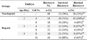
Table 1. Effect of developmental stage of biopsied embryos on cryotolerance of biopsy-derived blastocysts
a,b Data with different superscripts in the same column differ significantly (p<0.01)
|
|