TSGA10 is Specifically Expressed in Astrocyte and Over-expressed in Brain Tumors
-
 Behnam, Babak
M.D., Ph.D., Reproductive Biotechnology, Avicenna Research Institute, ACECR, Tehran, Iran, P.O. Box: 19615-1177, Tel: +98 21 22432020, Fax: +98 21 22432021, E-mail:b.behnam@avicenna.ac.ir;bbehnam@iums.ac.ir
Behnam, Babak
M.D., Ph.D., Reproductive Biotechnology, Avicenna Research Institute, ACECR, Tehran, Iran, P.O. Box: 19615-1177, Tel: +98 21 22432020, Fax: +98 21 22432021, E-mail:b.behnam@avicenna.ac.ir;bbehnam@iums.ac.ir
-
Department of Biology, University College London (UCL) , London, UK
-
Department of Microbiology and Molecular Biology, College of Medicine, University of Central Florida (UCF), Orlando , Florida, USA
-
Department of Genetics and Molecular Biology, College of Medicine, Iran University of Medical Sciences (IUMS) , Tehran, Iran
-
Chahlavi, Ali
-
St. Vincent's Spine and Brain Institute, Jacksonville , Florida, USA
-
Pattisapu, Jogi
-
Department of Microbiology and Molecular Biology, College of Medicine, University of Central Florida (UCF), Orlando , Florida, USA
-
Wolfe, Jonathan
-
Department of Biology, University College London (UCL) , London, UK
Abstract: In this study TSGA10 has been demonstrated as a testis-specific human gene that encodes a protein localized in sperm-tail and conserved in ciliary structure. Further investigations showed TSGA10 signalling and expression during embryogenesis, brain development and some malignancies including brain tumors. Given the role of this protein in neuronal development and in certain tumors, it could potentially serve as a diagnostic marker and therapeutic target in brain tumors. Therefore, using immunohistochemistry, we evaluated the localization of TSGA10 in different regions of brain, and its pattern/level of expression in tissue microarray (Cybrdi) containing human brain tumors and normal brain. In rat specimens, TSGA10 was mainly expressed in subventricular zone, hippocampus and granular layer of cerebellum of the brain. The antibody also stained the diverse and different types of human brain cancers. The TSGA10 was strongly over-expressed in glioblastoma and astrocytoma when compared to normal human brain. The expression of TSGA10 was also confirmed in astrocyte derived from a human astroctyoma cell line by immunocytochemistry. This study indicates that TSGA10 can be used as an immunohistochemical marker for human neuroglia and astrocyte cells and is over-expressed in brain tumors.
Introduction :
TSGA10 has been described as a 82-kDa protein expressed in testis and during devel-opmental stage of spermatogenesis and em-bryiogenesis (1). It has already been indicated that full-length TSGA10 is post-translational modified, and the processed N-terminus
27-kDa mature TSGA10 is located in the Fi-brous Sheath (FS) of sperm tail (2). However, the C-terminal 55-kDa part of TSGA10 accu-mulates in the midpiece of spermatozoa,
where it co-localizes with HIF-1a, and ex-pressed during brain development (3), and in some solid and nonsolid tumors as a testis-cancer antigen.
The solid tumors include melanoma, pan-creatic adenocarcinoma, hepatocellular car-cinoma (4) and cutaneous lumphoma (5), ovar-ian leiomyosarcoma, germ cell tumor and gas-tric adenocarcinoma (derived ESTs). The non-solid tumors include Acute Lymphocytic Leu-kemia (ALL) and Acute Myeloplastic Leuke-mia (AML) (6,7).
The TSGA10 interacts directly with hyp-oxia inducible factor-1-alpha (3) and contrib-utes to the ciliary structure; thus it may be-have such as VHL. In this study, we have shown the over-expression of TSGA10 in brain tumors, and our eventual goal is to determine its role in brain tumorigenesis.
Materials and Methods :
Immunohistochemistry
Result :
It has been shown that TSGA10 is ex-pressed in some particular regions of normal brain. The brain regions in which TSGA10 is mainly expressed include the granular layer of cerebellum, hippocampus and subventricular zone (Figure 1). This pattern of TSGA10 ex-pression in the brain suggested a possible high expression of TSGA10 in dividing astrocyte and/or glia cells.
This study has demonstrated TSGA10 over-expression in different pathologic types of human brain tumors with a significant differ-ence compared to normal brain. The signals were scored via a point system by two differ-ent reviewers and final scores were an aver-age of points in different samples by different reviewers.
The brain tumors include glioblastoma, astrocytoma (pilocytic, grades II and III), oligodendroglioma, and medulloblastoma with highest TSGA10 over-expression in grade II oligodendrogliom (average 5+ in three samples) followed by glioblastoma (Table 1, Figure 2 ).
Sixty three samples (dots) have been pro-vided in the tissue array slide with nine differ-ent histopathologic diagnoses including 8 normal brains (1+), 3 medulloblastoma (3+), 12 oligodendroglioma (3 grade II:5+, and 9 grade III:3+), 19 astrocytoma (9 grade II:4+ and 7 grade III:3.9+, and 3 pilocytic:4+), and 21 glioblastoma (4.2+). Medulloblastoma and oligodendroglioma (grade III) generally show
a lower expression of TSGA10 protein com-pared to other tumors. Although TSGA10 protein seems to be expressed more in grades II compared to grade III, this difference is minimal in astrocytoma whereas maximal in oligodendroglioma. Meanwhile, the highest expression of TSGA10 is observed in grade II oligodendroglioma. However, glioblastoma identifies maximum TSGA10 expression as a histopathologic entity among brain tumors (Table 1).
To investigate a possible expression of TSGA10 in an astrocyte, a cell line which was derived from a known human astrocytoma (grade III, WHO, as a gift) selected and endo-genous TSGA10 expression was shown within the cells. Using immunostaining and IF methods, the antibody against the C-terminal of TSGA10 could localize the protein as a single spot around the nucleus, and as several spots within the nucleus, respectively (Figure 3). We also found a significant increase in The TSGA10 expression in this human cell line which was derived from astrocytoma, utilizing protein analysis. Western blot shows a 82 kDa TSGA10 band which was more intense in above-mentioned cell line (astro-cyte derived from grade III astrocytoma) than the astrocyte cell line (A735), as verified by scanning densitometry normalized to ß-actin (Figure 4).
Discussion :
It has already been shown that TSGA10 protein has a post-translational modification, resulting in an N-terminus 27-kDa and a
C-terminus 55-kDa components (1). Although the C-terminus TSGA10 may be associated with an organelle such as centrosome which is consistent with its role in cell division and proliferation however a further study in tumor biology is necessary to confirm a precise
and specific function of each component.
In this study, we have demonstrated an over-expression of TSGA10 in brain tumors with a significant difference between normal brain and malignant cells. This is consistent with the previously reported over-expression in some other tumors (4-6; BLAST/NCBI ESTs). The TSGA10 (C-terminus) also inter-acts with HIF-1-alpha (3) and contributes to ciliary-centrosomal structure (1). The results of this study along with prior molecular findings on the TSGA10, may address a crucial role in regulating brain tumorigenesis, apoptosis and hypoxia pathways. These findings on the TSGA10 molecular behavior and expression also remind a similar function to VHL tumor suppresser protein.
Acknowledgement :
Authors would like to thank Prof. Sue Povey (UCL, London, UK) for providing the cell line astrocyte which was derived from a known human astrocytoma (grade III, WHO).
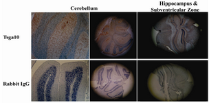
Figure 1. TSGA10 expression in rat brain
TSGA10 is expressed in different regions of rat brain including granular layer of cerebellum, hippocampus and Subventricular zone (SVZ). These brain zones are well known as the glia-rich regions of brain
|
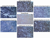
Figure 2. TSGA10 is expressed in human normal brain and over-expressed in human brain tumors
By immunohistochemistry and utilizing polyclonal antibody (raised in rabbit) and Cybrdi tissue microarray, TSGA10 expression is shown in normal brain, and in different brain tumors in human. TSGA10 expression shows a significant increase in human brain tumors compared to normal brain. �Glioblastoma� followed by �pilocytic astrocytoma� and �astrocytoma (grade II)� show the highest expression of TSGA10 protein and �medulloblastoma� the lowest among the brain tumors. (Zeiss microscope, x40)
|
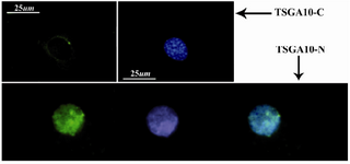
Figure 3. TSGA10 localization in astrocyte
In immunocytochemistry, TSGA10 antibody could detect the protein expression and localize the C-terminus (top panel) of the TSGA10 protein as a single perinuclear spot in the culture of astrocyte which is derived from human astrocytoma (WHO, grade III). However, the N-terminus of the protein (button panel) is expressed within the nucleus as several spots. DAPI used for the nuclei staining and FITC-conjugated (green) secondary antibody was used in IF
|
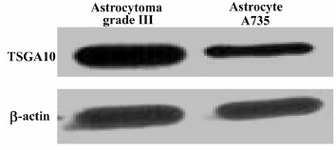
Figure 4. TSGA10 protein expression in astrocytes
Western blots for TSGA10 expression in astrocytes derived from a grade III astrocytoma (left panel) and an astrocyte cell line (735), in which its expression normalized to β-actin. TSGA10 protein levels in grade III astrocytoma is significantly increased compared to the astrocyte cell line (735).
|
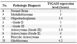
Table 1. TSGA10 protein expression in 63 human brain samples
|
|