miR-155 Down Regulation by LNA Inhibitor can Reduce Cell Growth and Proliferation in PC12 Cell Line
-
Kouhkan, Fatemeh
-
Department of Genetics, Faculty of Basic Sciences, Tarbiat Modares University, Tehran, Iran
-
Alizadeh, Shaban
-
Department of Hematology, Allied Medical School, Tehran University of Medical Sciences, Tehran, Iran
-
 Kaviani, Saeid
Saeid Kaviani - Ph.D., School of Medicine, Tarbiat Modares University, Tehran, Iran , Tel: +98 21 82883585 Fax: +98 21 82884555 E-mail: kavianis@modares.ac.ir
Kaviani, Saeid
Saeid Kaviani - Ph.D., School of Medicine, Tarbiat Modares University, Tehran, Iran , Tel: +98 21 82883585 Fax: +98 21 82884555 E-mail: kavianis@modares.ac.ir
-
Department of Hematology, School of Medicine, Tarbiat Modares University, Tehran, Iran
-
Soleimani, Masoud
Masoud Soleimani - Ph.D., School of Medicine, Tarbiat Modares University, Tehran, Iran, Tel: +98 21 82883585 Fax: +98 21 82884555 E-mail: soleim_m@modares.ac.ir
-
Department of Hematology, School of Medicine, Tarbiat Modares University, Tehran, Iran
-
Amirizadeh, Naser
-
Research Center of Iranian Blood Transfusion Organization (IBTO), Tehran, Iran
-
Abroun, Saeid
-
Department of Hematology, School of Medicine, Tarbiat Modares University, Tehran, Iran
-
Noruzinia, Mehrdad
-
Department of Hematology, School of Medicine, Tarbiat Modares University, Tehran, Iran
-
Mohamadi, Shahin
-
Department of Hematology, Allied Medical School, Tehran University of Medical Sciences, Tehran, Iran
Abstract: MicroRNAs (miRNAs) are a class of small non coding regulatory RNAs that have key functions in multiple cell processes. Deregulation of these tiny miRNAs are involved in various human diseases. MiR-155 is one of the multifunctional miRNA that its over-expression has been found to be associated with different kinds of cancer such as leukemia, breast and colon cancers. It is thought that deregulation and over-expression of this microRNA may be associated with PC12 cell proliferation. So, the aim of this study was to investigate the role of miR-155 expression on PC12 cell growth. For this reason, PC12 cells were cultured and transfected by 3 different concentration (25, 50 and 75 nmol) of either LNA anti-miR-155 or scramble antisense in 24-well plate. Then, total RNA was extracted from transfected cells. miRNA cDNAs were synthesized from isolated total RNA. In the second step, miR-155 expression level was analyzed using the quantitative real-time polymerase chain reaction (QRT-PCR). MTT test was performed to evaluate cell viability. In the next step, apoptosis assay was assessed to investigate anti miR-155 effect on PC12 cells death. Obtained results were analyzed with t-test. MTT test revealed that cell viability of transfected cells with 75 nM of anti-miR- 155 to be reduced by half of the control and scramble groups (0.5 vs. 0.97 and 0.94). Our data suggest that miR-155 over-expression is associated with PC12 cell growth. So, miR-155 down regulation by anti-miR-155 could open up new ways to restrain brain tumor growth, as anti-miR-155 causes PC12 cells to repress.
Introduction :
microRNAs are tiny regulatory molecules that have important role in various biological processes such as proliferation, cell death, dif-ferentiation and metabolism (1-5). Misregula-tion of these molecules is correlated with dif-ferent pathogenic situations including tumorogenesis (6). In cancer, miRNAs act as oncogenes or tumor suppressors. Over ex-pression of oncogenic miRNAs (oncomiRs) can down regulate tumor suppressors and/or other genes involved in cell differentiation, thus leading to tumor formation by stimulat-ing proliferation, angiogenesis, and invasion. Similarly, tumor suppressor miRNAs can down regulate different proteins with onco-genic activity (7-11). Recent studies point out that miR-155 frequently up regulates in vari-ous cancers, so it is considered as oncomiR (12).
Brain tumors are created by abnormal pro-liferation of cells that normally exist in the brain such as neurons or spread from cancers primarily located in other organs. However, any brain tumor is inherently dangerous and life-threatening. So, there is considerable in-terest in identifying more effective therapeutic strategies for treatment of brain tumors (10).
Anti-miRs are chemically modified, single stranded nucleic acids designed to specifically bind to and inhibit endogenous microRNA (miRNA) molecules. Thus, they are miRNA Inhibitors. In general, anti-miRs have many benefits in cancer treatment including effi-ciency, precision, versatility, speed and cost-ef¬fectiveness (13-19).
Therefore in this study, we investigated the anti miR-155 effect on repression of PC12 tumor cell growth and proliferation. The PC12 is a cell line that is established from rat pheochromocytoma. PC12 cells divide and resemble sympathetic neurons, when grown in Nerve Growth Factor (NGF) containing media. Thus, the PC12 cell line is used as a model system for neuronal cells.
Materials and Methods :
Cell cultures
Pc12 cell line was maintained in RPMI 1640 supplemented with 10% Fetal Bovine Serum (FBS) and 50 mg/ml penicillin/ strep-tomycin in a humidified incubator containing 5% CO2 at 37 C. Cells were fed three times a week and passaged every 7 days.
Oligonucleotide sequences
5' fluorescent antisenses: anti-155 (CCTA TCACGATTAGCATT, EXIQON, product no:410078-04);5'fluorescent scramble siRNA (GTGTAACACGTCTATACGCCCA,EXIQONproduct no:199004-08). QRT-PCR primers: miR-155 forward primer (5'-TTAATGCTA ATCGTGATAGG-3'); U6 forward primer (5'- CTCGCTTCGGCAGCACACATATAC-3'),U6 reverse primer (5'-ACGCTTCACGAATT TGC GTGTC-3').
Transfection and MTT experiments
Twenty four hr before transfection 5×10⁵ PC12 cells were seeded per well in 24 well plate and allowed to grow overnight. The cells were then transfected with three differ-ent concentration of anti-155 (25 nM, 50 nM and 75 nM, respectively) and highest concen-tration of scramble siRNA(75 nM) using lipo-fectamine 2000 (Invitrogene) according to the manufacture's protocol. Scramble siRNA dose not target any known miRNA in trans-criptome. These anti-miRs were purchased from Exiqon Company.
The day after transfection, medium was replaced with fresh RPMI containing 10% FBS. Cultures were incubated for the next day at 37 C. Then, the cells were treated with MTT solution (5 mg/ml) for 4 hr. Thereafter, Dimethyl Sulfoxide (DMSO) were added to each well and absorption at wavelength of 540 nm measured by a spectrophotometer.
miRNA extraction and real-time
Total RNAs were extracted from trans-fected cells using biozol reagent (Bioneer Company). miRNAs cDNA were synthesized by Stratagene kit according to manufacturer's protocol. Thereafter QRT-PCR of miR-155 was performed with general reverse primer of Stratagene kit and specific forward primer for miR-155. The miR-155 expression was nor-malized to an endogenouse control U6. Rela-tive expression was computed with relative standard curves for miR-155 and U6.
Apoptosis assay
Apoptosis assay was carried out in dupli-cate experiments using caspase-3 colorimetric activity assay kit (Millipore Company). Brief-ly, anti miR-155 transfections were performed as mentioned above. After 72 hr of trans-fection,0.5×106 cells were suspended in 100µl of chilled 1X cell lysis buffer and incubated on ice for 10 min and then centrifuged for
5 min at 10000 g. Assay mixtures were prepared in a 96-well plate. Twenty µl of 5X assay buffer and 10 µl of caspase-3 substrate (Ac-DEVD-pNA) were added to each test samples. Test samples were incubated for 2 hr at 37 C. Then absorbance at 405 nm was measured with a microtiter plate reader. Fold increase in caspase-3 activity was calculated by comparing the OD reading from the in-duced apoptotic test samples with the level of the un-induced control.
Statistical analysis
The t-test was performed to investigate the differences in the obtained results of the three groups in the MTT and apoptosis analysis. Probability of 5% was assumed statistically significant.
Results :
PC12 cells were divided in 3 groups: con-trol group, scramble group that was transfect-ed by 75 nM of scramble antisense and test group that was transfected by 25 nM, 50 nM and 75 nM of anti miR-155. Transfection efficiency was determined using inverted fluorescence microscope. Transfected cells with at least 70% efficiency were selected for further analysis. QRT-PCR analyses revealed that miR-155 expression was decreased in cells transected by different concentrations of anti-miR compared to control group (3 fold decrease for 25 nM, 5 fold decrease for 50 nM and 6.75 fold decrease for 75 nM concentra-tions of anti miR-155). No statically import-ant differences were observed between miR-155 expression in control and scramble groups (Figure 1).
In the next step, cytotoxic effect of dif-ferent concentration of anti miR-155 and scramble antisense were evaluated 48 hr after transfection by microscopy, trypan blue staining and MTT test. Optical densities at 540 nm were obtained for 3 groups: 0.971 for control group 0.962 for scramble group and 0.796, 0.659 and 0.561, respectively for
25 nM, 50 nM and 75 nM concentrations of anti miR-155 (Figure 2).
These results demonstrated that optical density and therefore cell viability was similar in control and scramble groups (t=3.60, p=0.06). However, there were significant differences between optical density and cell viability in anti miR-155 group compared to control groups (t=55.9723, p=0.0003, t=68.44, p=0.0002, t=182.01, p=0.0001, re-spectively for 25 nM, 50 nM and 75 nM con-centrations of anti miR-155).
Optical density and cell viability gradually decrease with increase in anti miR-155 con-centration (0.796, 0.659 and 0.561 respect-ively). At 75 nM concentration of anti miR-155, optical density and cell viability were half of these parameters in control group. These data indicated that higher concentration of anti miR-155 had higher toxicity effect on PC12 cells and could decrease the cell viabil-ity and proliferation.
In the next step, we tried to identify the toxic effect of anti miR-155 (due to its effect on apoptosis pathway). For this reason, cas-pase-3 activity was calculated in 3 groups by caspase-3 colorimetric activity assay kit. Optical densities at 405 nm were 0.099 for control group, 0.097 for scramble group and 0.117 and 0.119 for anti-miR-155 group, that respectively was transfected by 50 and 75 nM of anti-miR (Figure 3). According to these data, there were no statically differences be-tween optical density and caspase-3 activity between control and scramble groups (t=0.44, p=0.70) and control and anti miR-155 groups (t=1.34, p=0.31 for 50 nM concentration of anti miR-155 and t=1.32, p=0.31 for 75 nM concentration of anti miR-155).
Discussion :
microRNAs are a class of small non-coding single-stranded RNA molecules that partici-pate in control of gene expression by repres-sion protein translation or by destabilizing target mRNA via cleavage or deadenylation. Any changes in the expression level of spe-cific miRNAs are associated with multiple human diseases including diabetes, heart dis-eases, neurological diseases, hypertension and cancer (20-22).
Cancer is the multistep process caused by uncontrolled cell proliferation. Recent studies demonstrate that some miRNAs are consider-ed as oncogenes or tumor suppressors and have important roles in tumor initiation, pro-gression and metastasis (11,22).
In 2002 for the first times, Calin and his partners indicated that down regulation of miR-15 and miR-16 was linked with chronic lymphocytic leukemia. So, these genes are thought to function as tumor suppressor (23,24).
On the other hand, some miRNAs act as oncogenes and are called oncomirs. Up regulation of these miRNAs could cause the tumor progression (25). miR-155 can be regard-ed as an oncomir whose expression is increas-ed in different cancers including T cell lymphoma, leukemia, breast, thyroid and colon cancers (26-29). So, in this study, we utilized anti-miR-155 for repression of PC12 growth and proliferation. Transfection of PC12 cells with either anti miR-155 or scram-ble were done successfully by at least 70% efficiency. MTT test was performed and results indicated that although 25 nM and
50 nM of anti miR-155 have toxic effect, on PC12 cells, 75 nM of anti miR-155 could repress tumor cell proliferation strongly (half of the control group).
On the other hand, apoptosis analysis dem-onstrated that caspase-3 activity was similar in control group (0.099), scramble group (0.097) and anti miR group (0.117 for 50 nM and 0.119 for 75 nM of anti-155). Thus, anti miR-155 did not influence PC12 apoptosis. Consequently, anti miR-155 could decrease PC12 cells development, but could not induce apoptosis in these cells.
Previous studies have indicated that a num-ber of genes that are regulated by miR-155 are involved in biological processes such as immune response (for instance Pu1 and SHIP1), inflammation (BACH1 and ZIC3) and cell growth/survival (TP53NP1, FOXO3 and GSK3β) (12,26,30-35). Thus, miR-155 could promote cancer development and proliferation by targeting genes that control cell cycle arrest such as tumor protein 53-induced nuclear protein 1(TP53INP1), (25,31). However, there is little information from direct targets or exact pathways through which miR-155 signals function to promote the PC12 tumor cell development and proliferation.
Conclusion :
In summary, our data for the first time indicated that down regulation of miR-155 by anti-miRNA, that has sequence complemen-tary to mature miR-155, may be a useful approach for inactivating this microRNA in brain tumors and cause slow tumor growth, development and proliferation. Meanwhile further investigation would be necessary for identification of the exact mechanism through which anti miR-155 influence the PC12 tumor cells growth and proliferation.
Acknowledgement :
This study was accomplished at the Univer-sity of Tarbiat Modares and supported by the Hematology department. The authors are grateful to the department for their support.
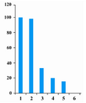
Figure 1. Relative Q-PCR of miR-155 level in PC12 cell line. (1) Control PC12 cells. (2) PC12 cells transfected by 75 nM of scramble. (3) PC12 cells transfected by 25 nM of anti miR-155(4) PC12 cells transfected by 50 nM of anti miR-155(5) PC12 cells transfected by 75 nM of anti miR-155
|
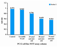
Figure 2. MTT assay on control, scramble and test groups
|
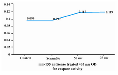
Figure 3. Effect of anti miR-155 on caspase-3 activity in control, scramble and test groups
|
|